The two strips and the valley between them
Two strips of cortex face each other across a deep fold that runs over the top of your head. The one in front ; the one behind . What you are turning above is the group-average human brain with those regions painted from the HCP-MMP1 atlas, and every click reports the atlas name, the correct hemisphere, and what that piece of brain does.
What the atlas is. HCP-MMP1 is the Human Connectome Project's multi-modal parcellation (Glasser et al., Nature 2016). It divides each hemisphere into 180 areas, 360 in all, drawn on the brains of 210 healthy young adults and then checked by finding the same borders again in a second, independent group of 210. An area earns a border only where several kinds of measurement change together: the myelin content of the cortex, its thickness, what it does during tasks, and which regions its activity fluctuates with at rest. The surface you are turning is the group average of those same brains, so the areas and the anatomy come from one consistent source. It is the parcellation working neuroscientists argue with, and about, today.
The fold is the central sulcus, and it is one of the few landmarks a brain reliably keeps. On its front bank sits area 4, primary motor cortex, whose giant output neurons run all the way to the spinal cord; most of their axons cross at the base of the brainstem, which is why each hemisphere moves the opposite side of the body. Watch a signal run the measured corticospinal tract. (The measured bundles descend to the midline at the medulla; the crossing itself is textbook anatomy that diffusion tractography cannot cleanly trace.) On the back bank, primary somatosensory cortex is not one area but four narrow strips in a row, , , , , and the order matters: signals about muscle stretch arrive deepest in the fold, skin touch lands just behind it, and each strip further back builds something more assembled, from raw contact to texture to the shape of the thing in your hand.
Around those two strips the selection widens: planning movements before M1 fires, the where self-initiated action starts, the , and keeping track of where your limbs are. The Areas control walks through them, and every card says which is which.
The little man this page refuses to draw
The textbook picture of this cortex is the homunculus: a distorted figure draped along the strip, feet on the medial wall, face at the bottom, hands enormous. The broad gradient is real: toes really are represented near the top of the head and lips near the ears, and the hand really does take an outsized share. But the tidy little man is a 1950s summary of stimulation surgeries, and modern measurements keep politely ruining him.
Precision imaging has found a somato-cognitive action network whose regions alternate with the foot, hand and mouth territories, so the motor strip is not one continuous body but an interleaving of body-part-specific and whole-body regions (Gordon et al., Nature 2023).
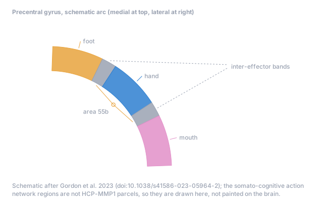
Receptive-field mapping shows body parts overlapping rather than parked in fenced plots, more broadly in M1 than in S1 (Schellekens et al., 2018). And intracortical recordings across the precentral gyrus found information about the whole body at every site sampled, arranged as biases in a mosaic rather than islands, with the honest caveat that those arrays mostly sampled the gyral crown, which is largely premotor territory (Deo et al., Nature 2026).
So this page paints what the atlas actually claims, where the areas are, and keeps the homunculus as history. And the measured body map is now here: switch Measured map on above and the surface wears the responses of 62 adults moving toes, ankles, legs, fingers, wrists, arms, jaws, lips, tongues and eyes in a scanner (OpenNeuro ds004044, CC0), with overlap and uncertainty shown rather than smoothed away. Washed-out colour means the winning movement barely beat the runner-up; the Stability view shows where individuals disagree with the group. It is labelled as the movement map it is.
What an atlas is, and is not
The parcellation here is HCP-MMP1: 180 areas per hemisphere, drawn where cortical architecture, function, connectivity and topography all agree a border belongs, in 210 healthy adults (Glasser et al., Nature 2016). It is the standard reference map of the human cortex, and it is a population map. Your area 4 is not exactly this area 4: individual brains differ enough that surgeons map each one afresh, with the patient awake and answering. An atlas tells you what is generally true about everyone; it cannot tell you what is precisely true about you.
Reference guide: the sensorimotor cortex
1. The landmark. The central sulcus is one of the few folds every human brain reliably has. Everything in this page is organised around it: motor cortex on the anterior bank, somatosensory cortex on the posterior bank, and a body map draped along both, feet at the top of the head near the midline, face down by the ear, hand claiming the largest share.
2. Output. Area 4, primary motor cortex, contains the giant layer 5 pyramidal neurons whose axons form the corticospinal tract, about 134 millimetres of measured cable from cortex to cord in this site's tract atlas. Most fibres cross at the pyramidal decussation, so each hemisphere moves the opposite body. M1 does not act alone: dorsal and ventral premotor cortex select and shape movements, the supplementary motor area on the medial wall handles self-initiated sequences, and the cingulate motor areas bring motivation and effort into action, with their own spinal projections.
3. Input. S1 is four strips in functional order. Area 3a, deepest in the sulcus, receives proprioception from muscle spindles and joints. Area 3b, the classical primary touch map, receives fine cutaneous input. Area 1, on the crown, builds texture and motion across the skin. Area 2, rearmost, combines touch with position into shape and grasp. Behind them, area 5 integrates limb state for action; below, S2 and the parietal operculum hold second-order body maps that receive from both body sides; area 43 takes the mouth and tongue territory where the sulcus meets the operculum.
4. The insula. Buried in the Sylvian fissure, the insula carries the body's internal state: its posterior granular cortex is primary interoceptive territory for temperature, pain-related and visceral signals, and its anterior divisions integrate that state with emotion and salience. In 2026, intracranial recording and stimulation showed the insula also encodes a somatotopic motor map with effector-specific connections to M1 (PNAS, doi:10.1073/pnas.2517734123). The body is represented in more than one place.
5. The homunculus, then and now. Penfield's cartoon summarised stimulation surgeries: a single distorted body along the strip. The broad gradient is real. But precision imaging found a somato-cognitive action network interleaved with the foot, hand and mouth territories (Gordon et al., Nature 2023, doi:10.1038/s41586-023-05964-2), population receptive fields overlap rather than parcel the body (Schellekens et al., NeuroImage 2018, doi:10.1016/j.neuroimage.2018.06.062), and intracortical recordings across the precentral gyrus found whole-body information at every sampled site, arranged as a mosaic of biases, sampled mostly on the premotor crown (Deo et al., Nature 2026, doi:10.1038/s41586-026-10653-x). The strip is a real map and not a tidy one.
6. The atlas. The parcellation painted here is HCP-MMP1: 180 areas per hemisphere, drawn where architecture, function, connectivity and topography agree in 210 adults (Glasser et al., Nature 2016, doi:10.1038/nature18933). The Every area mode paints all 360, and hovering or tapping names any of them. It is a population reference: individual brains differ enough that surgical mapping is still done awake, person by person. The measured whole-body movement map from OpenNeuro ds004044 (62 adults, CC0) now ships on this page as the Measured map control, with its overlap and uncertainty shown rather than smoothed away.
This is a population-level educational map built from a group-average atlas. Individual brains vary, body representations overlap, and nothing here is a diagnostic or surgical map.