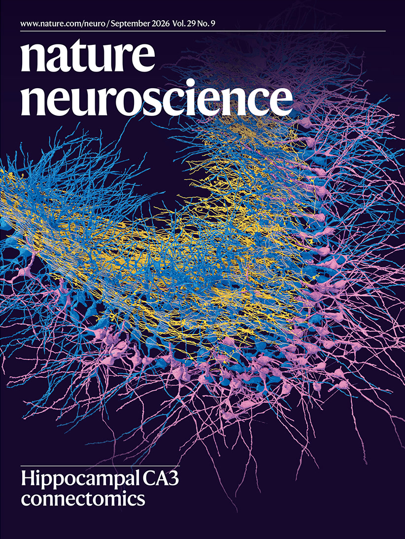
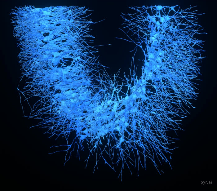
Every image on this page shows real cells from the CA3
region of the mouse hippocampus. First findings and a resource overview were
published in September 2026 in Nature Neuroscience.
The block that was sectioned, imaged and reconstructed: 1 x 1 x 0.1 mm
of mouse CA3, live. Drag to turn it. One pyramidal cell is drawn inside
at true relative scale. Nearly two thousand of them are in there, threaded through
by more than fifty five thousand mossy fibers. Pinch or scroll to zoom, or open
the full screen version.
Overnight renders
Rendered on one workstation in a single night, 103 minutes of GPU time for
the four pieces below. The two widescreen masters also replace their vertical
cuts further down this page when you are on a wide screen.
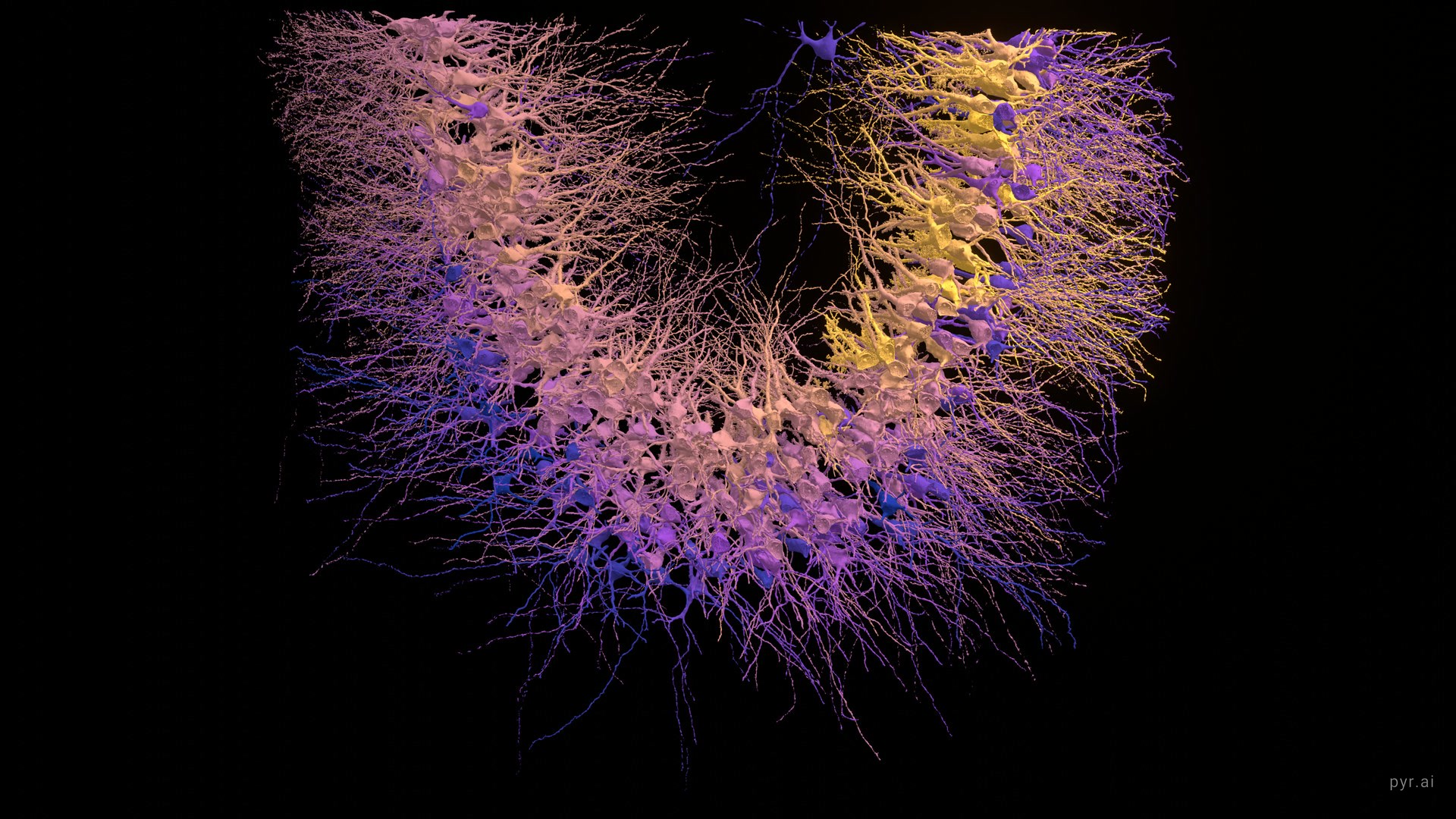
The convergence gradient. 304 pyramidal cells coloured by how
many mossy fibers reach them, deep blue for the fewest and gold for the most, on a
log scale because the count runs from 0 to 235 with a median of 41. Every cell here
sits in the deepest part of the block, where the reconstruction is most complete.
Read along the curve of the cell layer and the gradient is not a simple ramp:
both ends carry heavily innervated cells and the middle carries the fewest. In this
sample the median runs about 50 at one end, dips to 33 mid curve, then climbs to 72
at the far end, where the busiest cells reach 160 and above.Selective feedforward inhibition. Eighteen seconds. Seven mossy
fibers converge on one interneuron, in emerald, and its axon then makes 333
synapses onto 87 thorny pyramidal cells against only 50 onto 21
sparsely thorny ones. Normalised by how many cells of each subtype exist here, that
is 2.49 times more inhibition landing on each thorny cell. The wave travels
the interneuron's own cable, and each target lights at its real path distance, from
216 to 734 µm out.The scale ladder. Twenty seven seconds from the whole block to
a single synapse, holding on each rung long enough to read it. The tissue fills
with 1,064 cells, the view closes on one pyramidal neuron, then on the one thorny
excrescence where a mossy fiber wraps around it and makes 53 synapses inside
a box under seven micrometres on a side, and the spike arrives. Registration was
checked independently: this cell's soma lands on the stratum pyramidale boundary in
the real EM, with those boutons 39 um into the mossy fiber band.The gradient, measured as it goes. A band of light travels the
cell layer and the readout recomputes the median mossy fiber input count for the
cells inside it, from the same measurements that set the colours, so the number
and the cells lit under it cannot disagree. Watch the figure rather than the
caption: it starts near 51, falls to 33 through the middle, and
climbs past 64 at the far limb, where the busiest cells carry more than
170. The count of cells in the band is shown throughout, because the
sample thins at the ends and a median over twenty cells deserves less weight
than one over eighty.
Why the variation matters
The textbook picture of this connection is a detonator. Each CA3 pyramidal
cell is supposed to hear from only a handful of granule cells, but so powerfully that
one of them can nearly fire the cell by itself. Few inputs, each enormous, is exactly
the arrangement you would design if you wanted two similar experiences to end up
recruiting two different sets of CA3 cells — which is what pattern
separation means, and why this synapse is thought to matter for laying down a
memory that does not smear into yesterday's.
That picture assumes every CA3 cell is roughly alike. This gradient says otherwise.
How much dentate input a cell receives depends substantially on where it sits
along the layer, and the spread is not subtle: the busiest cells here carry more than
five times the median of the quietest stretch. A cell's position is part of its job
description, so CA3 is less a uniform pool of interchangeable units than the classical
model treats it as.
The most interesting part is the twist, and it cuts against the simple reading.
The distal cells that collect the most inputs collect them through
smaller terminals holding fewer vesicles. Count and per-contact strength
run in opposite directions. So the gradient in the numbers above is not a gradient in
raw drive, and reading it as one would overstate it — a cell with three times
the contacts is not being shouted at three times as loudly. It looks more like the
same total budget being spent two different ways: a few loud inputs at one end of the
layer, many quiet ones at the other.
The wiring also turns out to be spatial. Cells here share far more mossy
fiber inputs than chance allows, but that sharing is explained better by which cells
sit near each other than by how many inputs each one has. Neighbours sample the same
passing fibers. That is in real tension with pattern separation, which works best when
nearby cells hear different things, and it is the kind of constraint you only see once
you can trace every fiber rather than sample a few.
What this cannot tell you. Terminal size and vesicle count
are anatomical proxies for synaptic strength, not recordings of it; nobody
measured a current here. The gradient is traced across roughly 1.1 mm of one
block from one animal, which is a slice of the proximodistal axis rather than the
whole of it, and one animal cannot tell you what varies between animals. And the
sample thins toward both ends, which is why the cell count sits in the readout the
whole way along.
The population, widescreen. Twenty seconds. Populations arrive
in circuit order: mossy fibers first, then the cells they drive, then the wider
pyramidal population, and inhibitory interneurons last.The synapse story, widescreen. Nineteen seconds. The same
sequence as further down the page, rendered for a wide screen rather than
cropped to one.
These two are tall subjects in a wide frame, so they carry black at
the sides. The population is deeper than it is broad and nothing is cropped to hide
that.
Watch it assemble
Populations arrive in circuit order: mossy fibers first as the incoming
signal, then the CA3 cells they drive, then the wider pyramidal population, and
inhibitory interneurons last.
What the study found
The hippocampus is where experience becomes memory, and CA3 is the part that binds
the pieces of an event together. To understand how, you need to know not just which
cells are present but exactly which ones talk to each other.
This study took a block of mouse CA3, imaged it with an electron microscope, and
traced every neuron and every connection inside it. The result is a complete wiring
diagram of a small piece of real brain: 1,815 pyramidal cells, the main
excitatory neurons, 229 inhibitory cells, and more than 55,000 mossy
fibers, the axons carrying signals in from the neighbouring dentate gyrus.
Not all pyramidal cells are alike
Some are covered in elaborate spiny structures called thorny excrescences, and these
cells receive many mossy fiber inputs. Others, the sparsely thorny cells, receive almost
none. The split is sharp rather than gradual, so these are genuinely two kinds of cell
rather than two ends of one continuum.
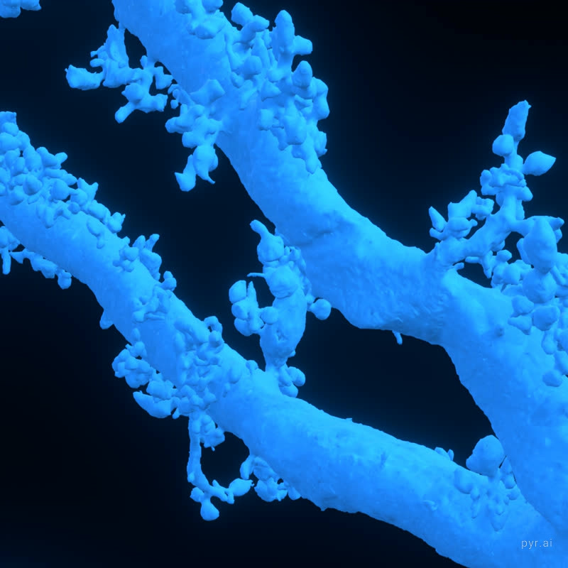
Thorny excrescences on pyramidal cell bodies. Each balloon like
cluster is a single elaborate spine, and a mossy fiber terminal wraps around it.
Input is unevenly spread across space
Cells further along the region receive substantially more mossy fiber contacts than
cells nearer the beginning. Cells sharing the same incoming fibers also sit closer
together than chance would predict, suggesting the wiring is organised rather than
arbitrary. Curiously, the cells receiving the most inputs get them through
smaller terminals holding fewer vesicles, so more connections does not simply
mean more of everything.
Inhibition is targeted, not general
Mossy fibers also drive inhibitory neurons, which act as brakes on the circuit. But
they drive only the inhibitory neurons that go on to target thorny cells. Those serving
sparsely thorny cells receive no mossy fiber input at all. So the same signal that
excites one type of cell also recruits the brake belonging specifically to that type,
leaving the other type untouched.
182 thorny pyramidal cells
Inside a single cell
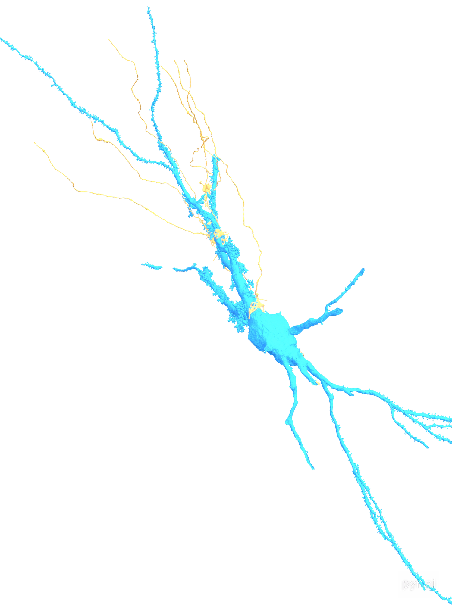
Thorny excrescences are the elaborate spines that give thorny pyramidal cells their
name. A mossy fiber ends on one in a single enormous terminal, making dozens of contacts
at one site rather than one contact in many places. This cell receives
165 mossy fiber synapses from just 6 fibers.
this one cell receives
165
mossy fiber synapses, and they arrive from only 6 fibers.
A single terminal makes dozens of contacts on one thorn.
reconstructed at
4.5M
triangles, with no simplification applied, which is why the
thorny excrescences hold their shape this close up.
in the full volume
1,815
pyramidal cells, 229 inhibitory cells and more than
55,000 mossy fibers were reconstructed.
Eight seconds, thorns to whole cell. The mossy fibers arrive
nearest first, so the one already in frame is the one that appears.
Watch it fire
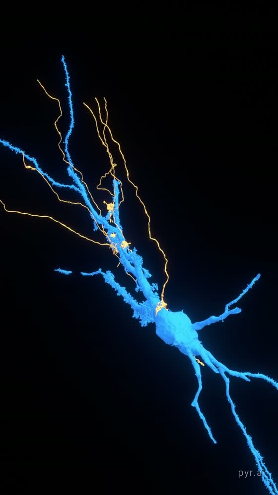
Six mossy fibers reach this cell, and this is the honest version of what they do.
One fires, lands hard on the thorn, and the cell does not answer. The other five
arrive. All six fire together, and only then does the cell send a signal of its own.
The pulse is not a sweep across the screen: every point on these meshes carries its
own distance measured along the cell's own skeleton, so the wavefront follows the
real branching and splits wherever the cable splits.
Eighteen seconds. All 165 mossy fiber synapses onto this one
cell, from all six fibers that reach it.
A single mossy fiber makes one of the strongest connections in the
brain, touching a CA3 pyramidal cell at dozens of release sites at once. Even so, one
incoming spike is usually not enough to make the cell fire. When the granule cell
fires a rapid burst of a few spikes, the connection strengthens within milliseconds
and then reliably triggers a spike. That is why these are called
conditional detonator synapses.
Two limits worth stating. The reconstruction is cut off at the edge of the
tissue block, so this arbor is incomplete. And the descending branches carrying the
signal are mostly basal dendrite with the axon embedded in them; at this resolution
the two could not be cleanly separated.
Three other cuts of the same event are here.
Every synapse these cells make
Sixteen CA3 pyramidal cells were traced. Fifteen of them have their axon followed
through the volume, and those axons make 25,723 synapses onto 6,348 other
cells in the same block of tissue. Every one sits at its real measured coordinate, not
an estimated position.
That works out to a median of 1,949 outgoing synapses per cell,
which sounds impossibly high until you remember what CA3 is for. Its pyramidal cells are
the most heavily interconnected population in the hippocampus, wiring back onto each
other through vast recurrent axon collaterals, and this is the network that theory says
lets the hippocampus complete a whole memory from a fragment of one. The gold beads
string along each axon because that is physically where the contacts are made, one
after another as the axon passes its targets. Most partners are touched just once:
58% receive a single synapse.
The presynaptic cells fade up, their synapses bloom across the volume, and then the postsynaptic partners arrive around them.
A sea of mossy fibers
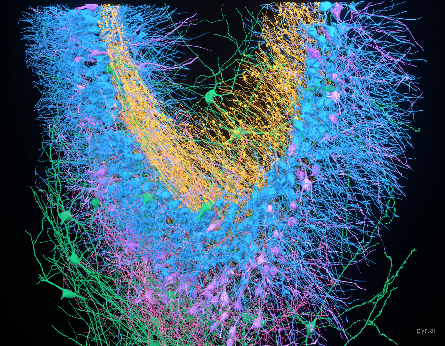
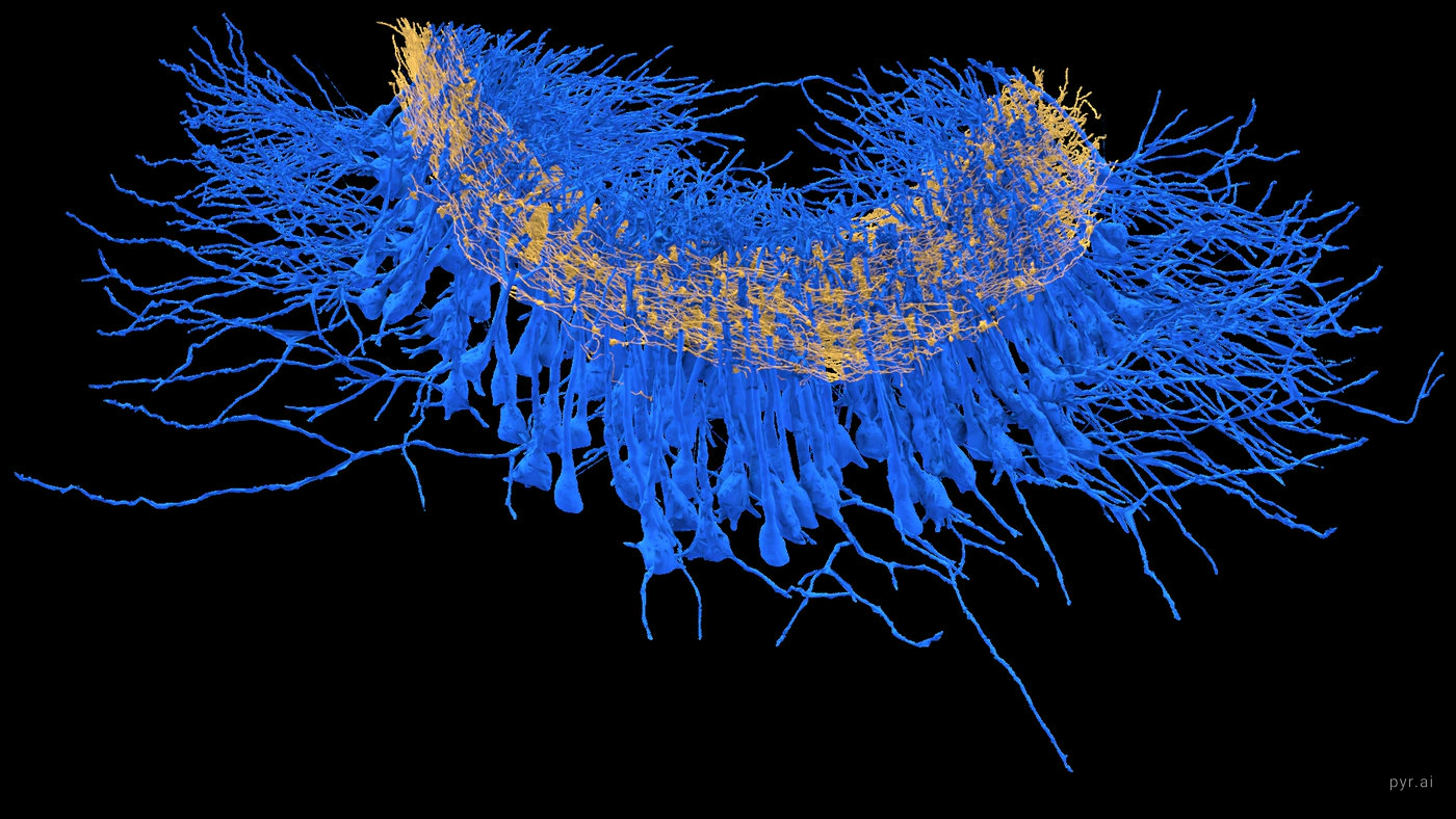
Seen edge on, the pyramidal cell bodies hang in a single layer while the mossy
fibers thread horizontally through them. Every gold strand is an axon arriving from
the dentate gyrus.
The classification, reproduced
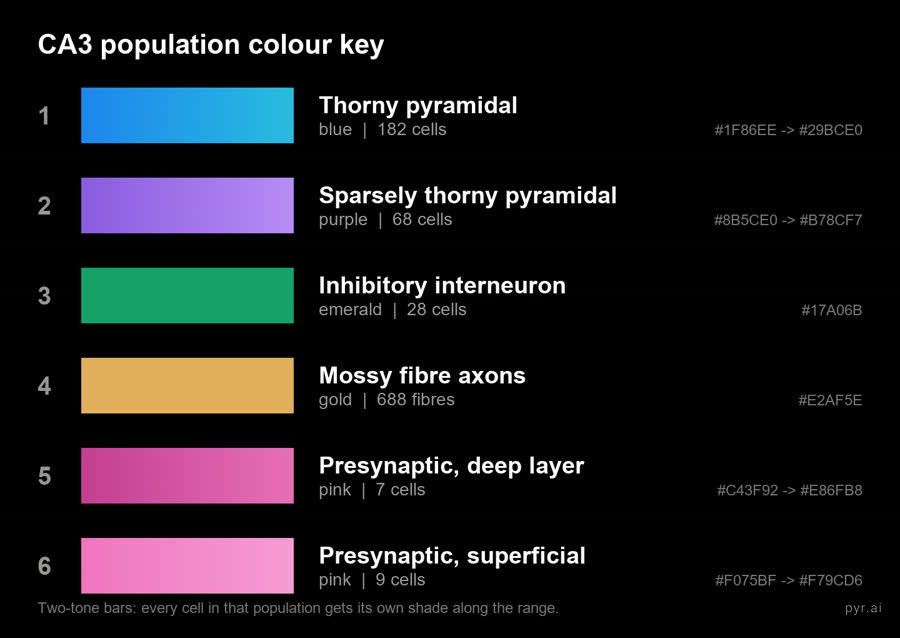
The study separates thorny from sparsely thorny cells by mossy fiber convergence,
drawing the line at more than 10 inputs. That split reproduces cleanly in this subset.
population
with MF input
median inputs
above 10
Thorny pyramidal
94 / 182
22
71%
Sparsely thorny
8 / 68
1
0%
Inhibitory
9 / 28
2
22%
Thorny cells reach 165 mossy fiber inputs at the top end.
Sparsely thorny cells max out at 3, and not one crosses the threshold. Only
9 of 28 interneurons receive mossy fiber input at all, which is the selectivity
the feedforward inhibition result describes.
Gallery
Stills from the same reconstruction, at full size, and one slow pass across the
convergence gradient. Tap any still to open the large version.
A slow pass across the gradient. Twelve seconds, a sixth of a
turn, eased at both ends so the arch is read rather than spun. Colour is how many
mossy fibers reach each cell, the same measurement as the still further up. What
the movement adds is depth: these are 304 cells occupying a curved sheet of
tissue, not points on a scatter plot.
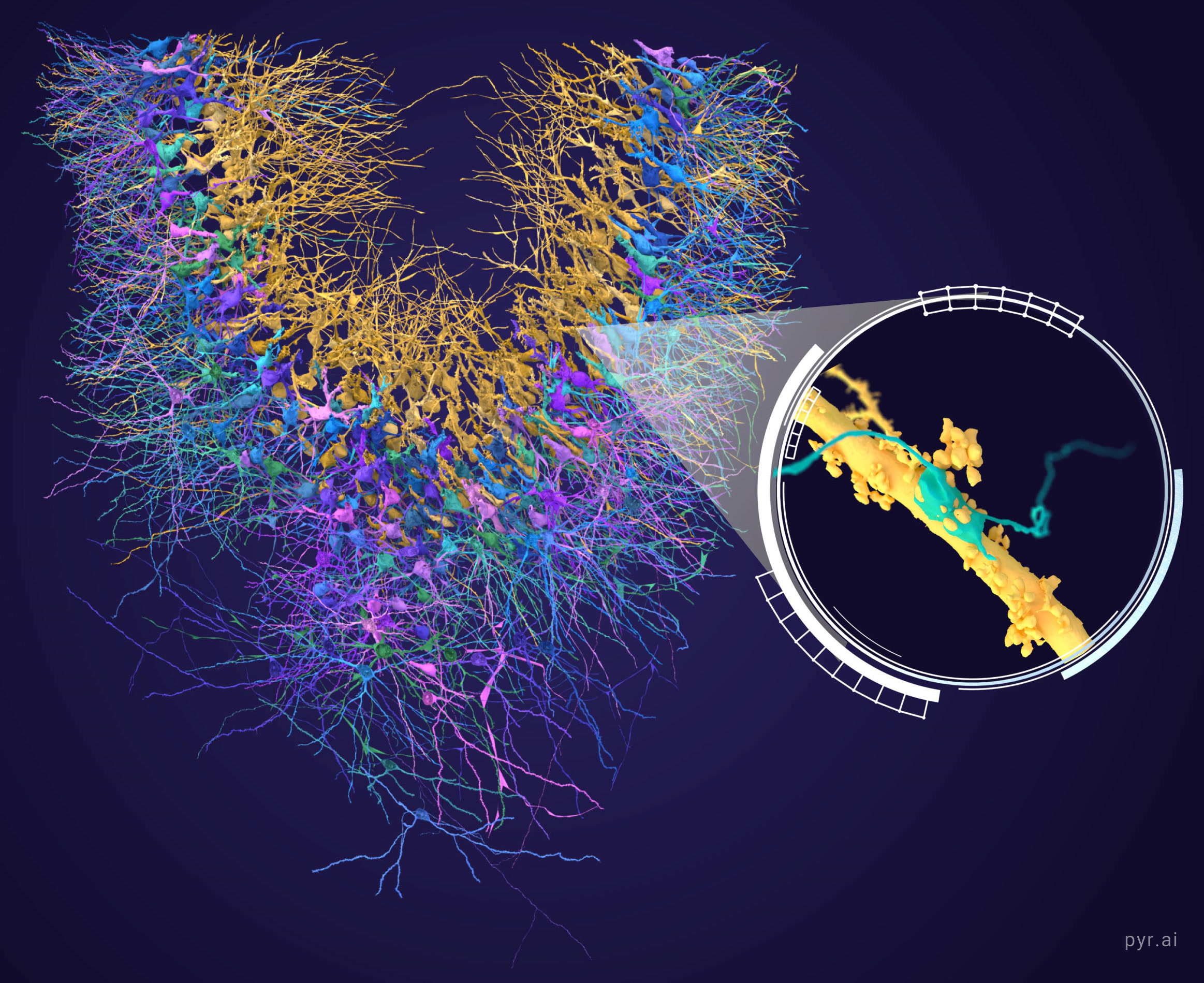
The reconstructed population seen face on, gold mossy fibers threading
through crowds of cells. The inset follows one mossy fiber axon down to its boutons,
with a partner cell process pressed against it.
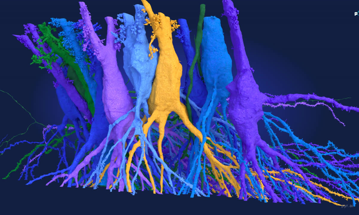
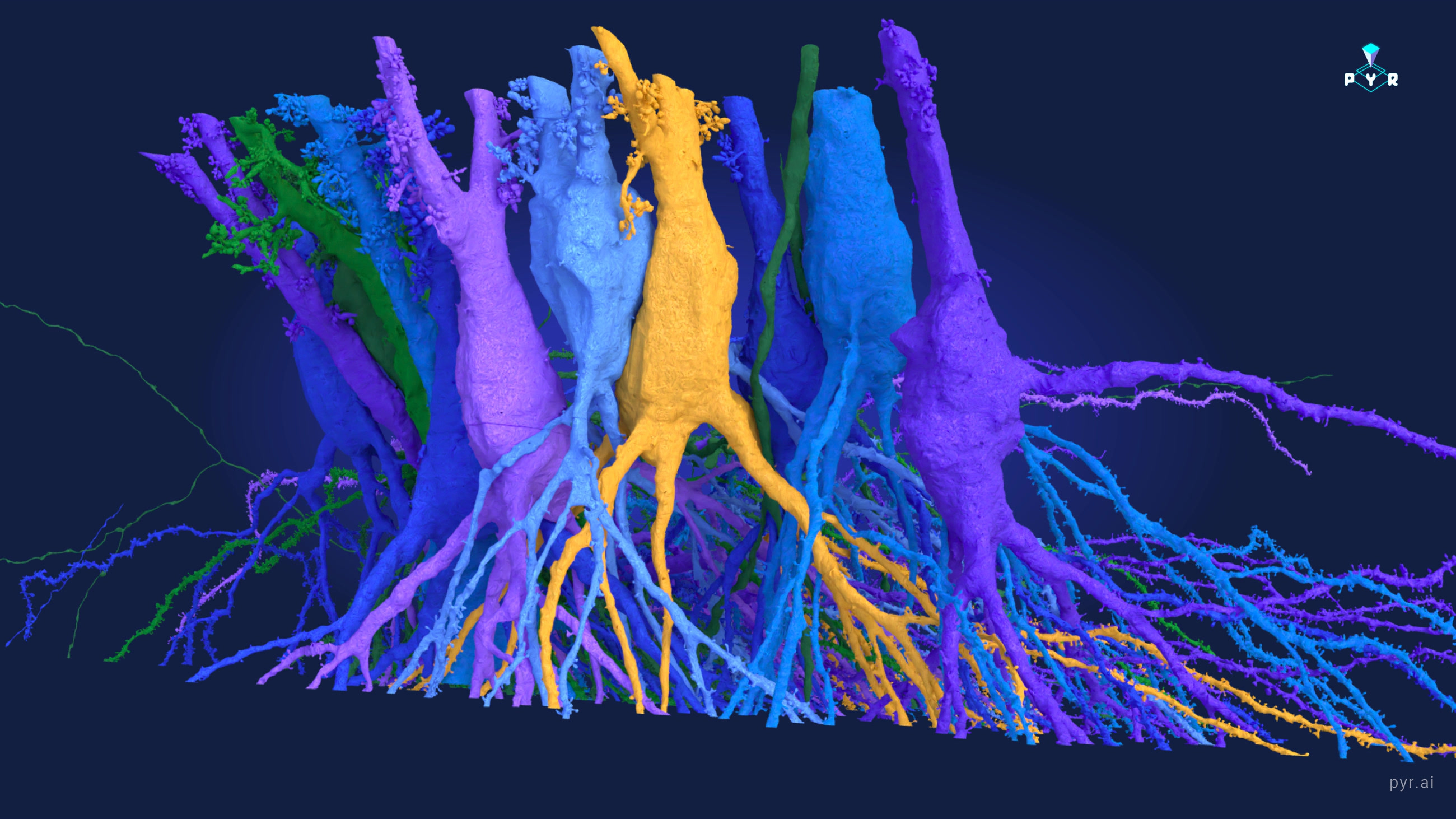
Pyramidal cell bodies lined up as they sit in the layer, one colour each.
The apical dendrites climb out of the top carrying their thorny excrescences, and the
basal dendrites fan out below.These six mossy fibers are not private to this cell. Each carries on
through the tissue and lands on 56 further segments, another 126 synapses.
Fourteen of those are complete cells with a nucleus in the volume, split evenly
between the two pyramidal subtypes; the rest are traced fragments. The purple is
not senders, it is fellow listeners hearing the same signal at the same moment.
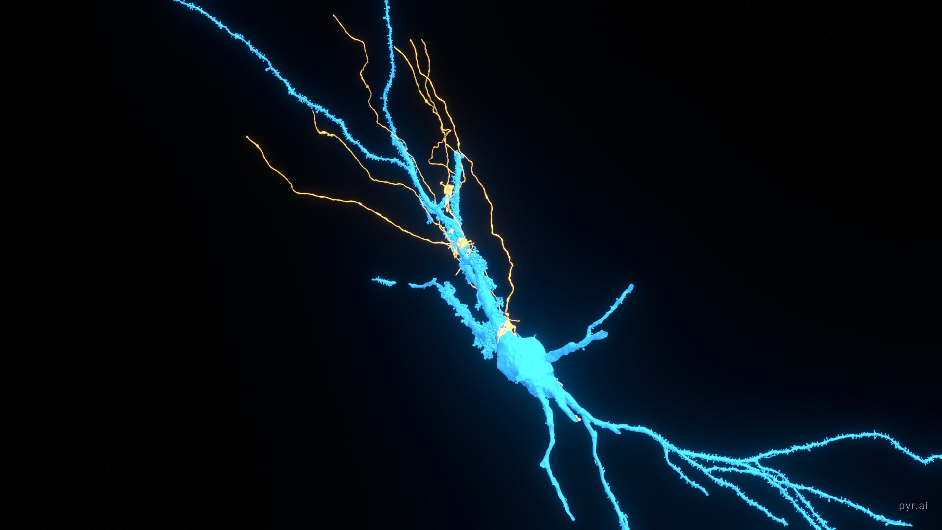
One thorny pyramidal cell and the six mossy fibers that reach it.
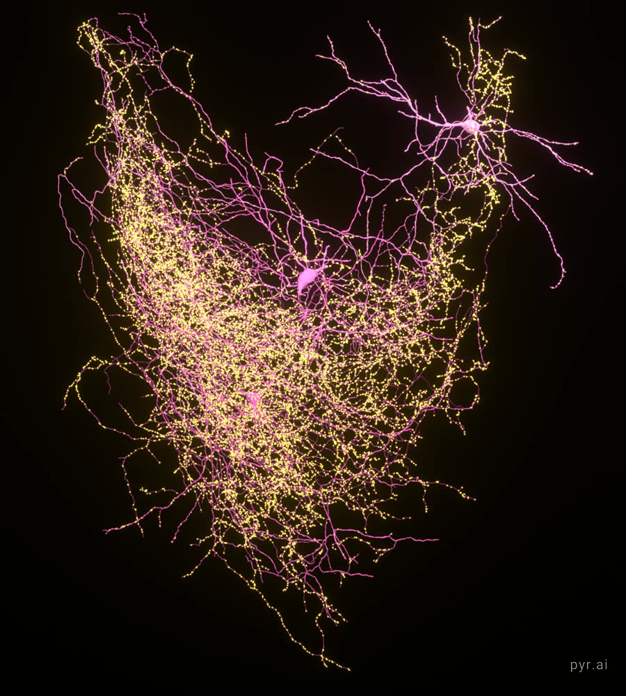
Sixteen presynaptic cells in pink, with all 25,723 of their synapses
in gold. The beads string along each axon because that is physically where the
contacts are made, one after another as it passes its targets.
A thorny excrescence up close. This tangle is a single spine.
A mossy fiber terminal in gold, wrapped around a thorny excrescence.
The blue thorns push out through the terminal that engulfs them.