microns mouse visual cortexa second dataset
Cortex
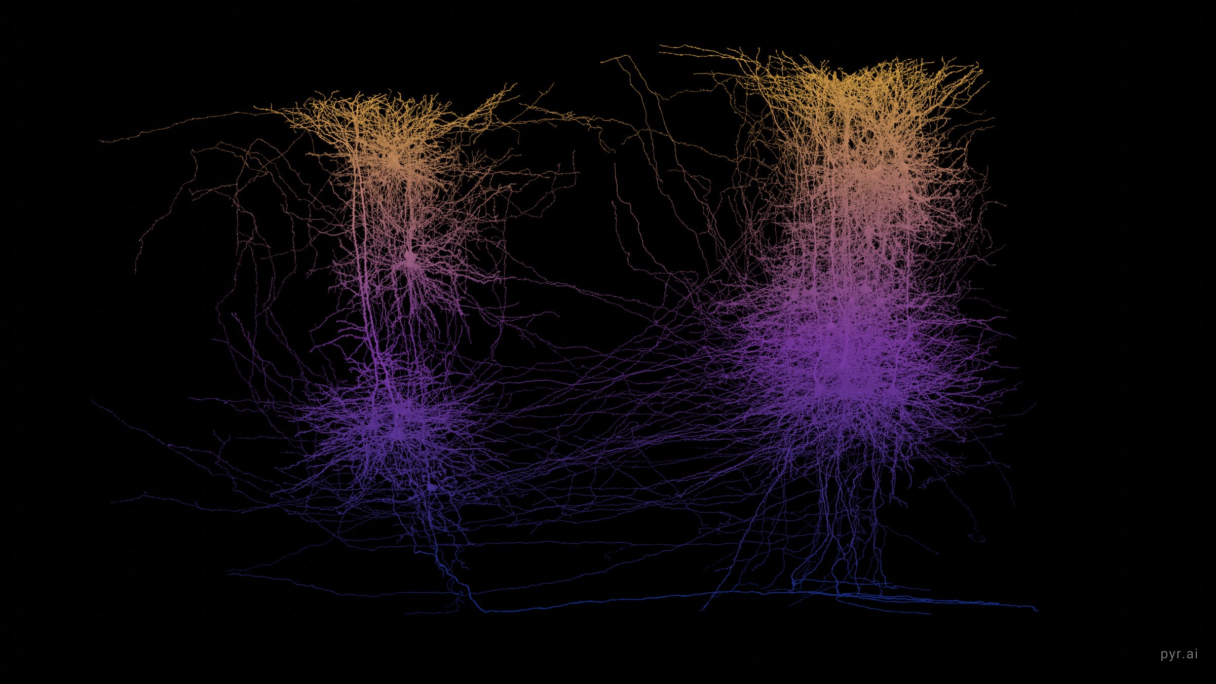
The same measured geometry, a different piece of brain. These are proofread neurons from the MICrONS volume, coloured by how deep they sit.
microns mouse visual cortexa second dataset
The same measured geometry, a different piece of brain. These are proofread neurons from the MICrONS volume, coloured by how deep they sit.
Every fully proofread neuron I hold from the MICrONS mouse visual cortex volume, 38 of them, seen side on with the pia at the top. Colour is not a property of the cell. It is position: every point on every arbor is tinted by its own depth below the surface, so one pyramidal neuron reads gold where its apical tuft reaches layer 1 and blue where its soma and basal dendrites sit deep. Nothing draws the layers in. They appear because that is where the cell bodies are.
the ramp spans
310 – 1083 µm
below the pia. Gold at the top of that range, deep blue at the bottom, violet through the middle. Every point on every arbor is coloured by its own depth, so one neuron carries the whole ramp along its length.
located cell bodies
34 of 38
span 429 to 910 µm, median 630, which is layer 2/3 through layer 6. Four cells have no nucleus in the table and carry no marker.
The same cells, moving. A still of a column is ambiguous in one specific way: you cannot tell whether the cells occupy a slab of tissue or happen to line up from one viewpoint. The only way to answer that is to move the camera.
These cells were chosen because they are proofread, not because they are a sample of anything. A neuron earns that status by a person checking it, so this is a picture of 38 well traced cells, not of a cortical column. Real cortex at this depth holds tens of thousands of neurons in the same space.
Nine of the mesh identifiers had gone stale since they were downloaded, because proofreading keeps merging and splitting segments, and were resolved forward through the chunked graph before the somata were looked up. Four cells have no nucleus in the table and carry no soma marker.
minnie65_phase3_v1.
To keep a clip, long press it on a phone or right click it on a desktop.